Nowadays, it is important not only to provide a high-quality result of dental rehabilitation of the patient but also to reduce the number of necessary visits to the minimum possible.
The patient applied to the dental clinic for consultation regarding the possibilities of correcting her smile.
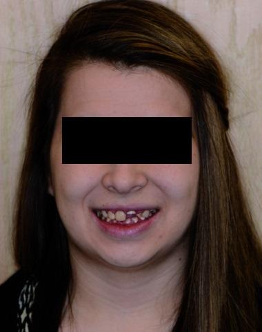
The patient photo before starting the treatment.
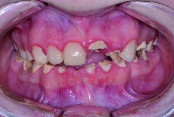
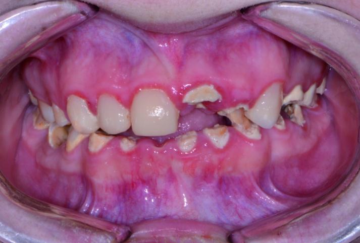
The appearance of the patient's teeth before starting the treatment.
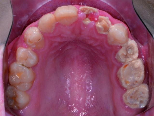
An occlusal view of the upper jaw before starting the treatment.
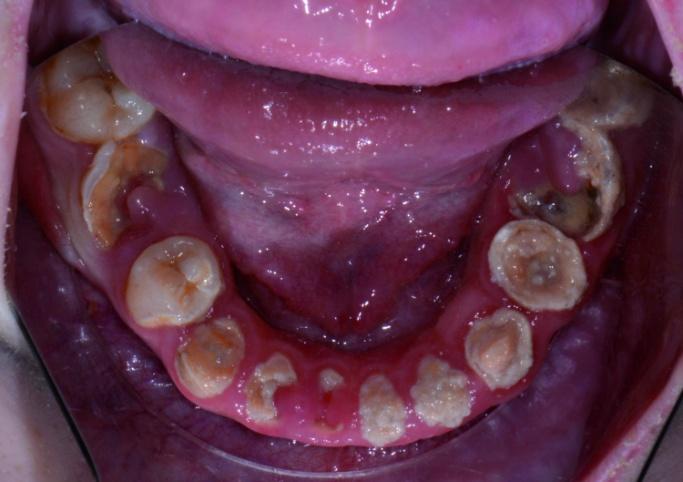
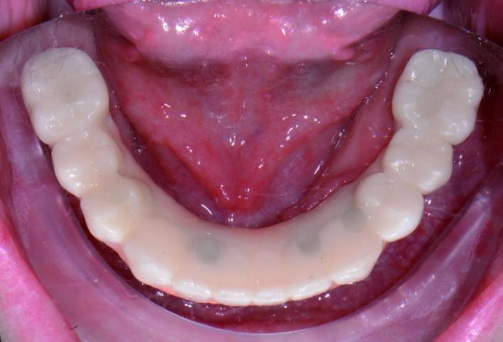
An occlusal view of the lower jaw before starting the treatment.
The process of treatment assumed the extraction of teeth No. 2-15 and No. 18-31, as well as of impacted third molars (teeth No. 1, 16, 17, 32).
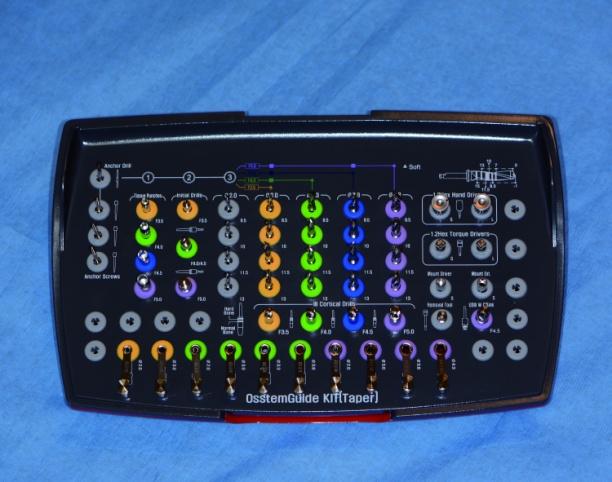
After analyzing all possible ways of treatment options, it was chosen a fixing all-arc zirconium prosthesis structures based on six dental implants on both the upper and lower jaws. In this clinical case, the implant system ET III SA (Hiossen) was used.
After removing all the teeth in the upper jaw the alveolar ridge of the upper jaw was aligned 2-3 mm in the apical direction. This procedure helps to hide the area of the transition of the prosthetic structure into the soft tissues of the oral cavity when the patient smiles.
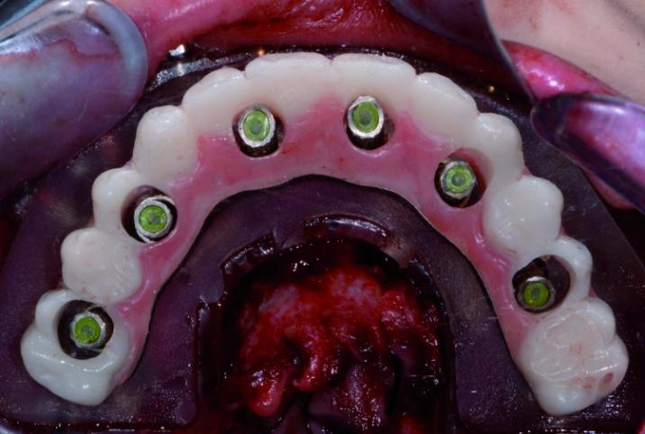
Dental implants ET III SA with a diameter of 4.0 mm were installed on the upper jaw in the area of teeth No. 4, 6, 8, 9, 11 and 13. Thus, the ‘All-on-6’ protocol was essentially implemented on the upper jaw. Implants of different diameters (3.5, 4.5 and 5.0 mm) were used on the lower jaw, taking into account the different width of the residual ridge in different anatomical areas.
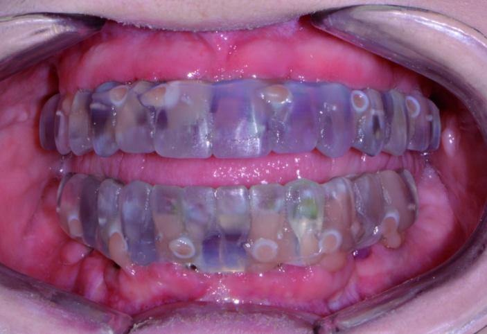
Provisional restorations were fixed on temporary abutments.
The patient was very happy with her temporary prostheses.
After 4-5 months, impressions were obtained for the manufacture of the final prosthetic restorations. About 16 weeks after implantation, the patient returned to complete the orthopedic phase of treatment.
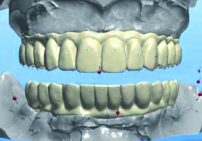
The duplication of the provisional restorations.
Planning the prosthesis design in 3Shape.
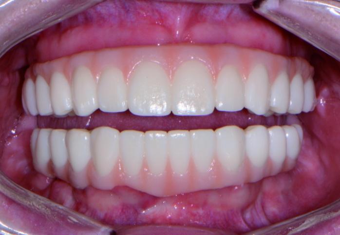
Permanent monolithic zirconium restorations have been successfully installed.

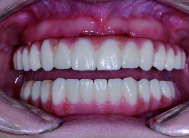
The view after fixing the final restorations.
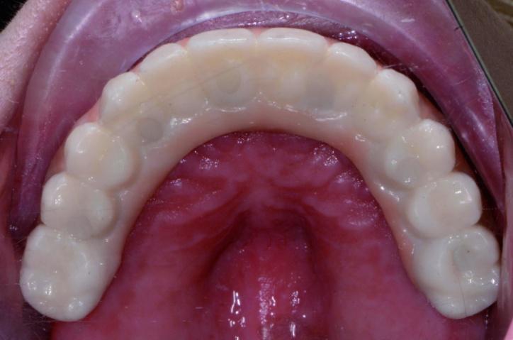
Occlusive view of the upper jaw after treatment.
Occlusive view of the lower jaw after treatment.
The final photo after the treatment.


